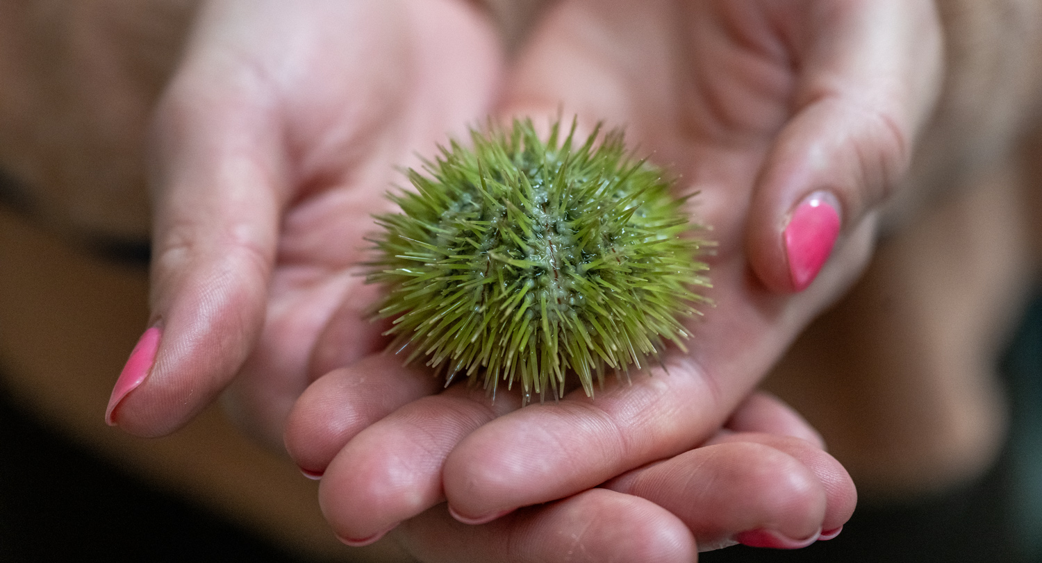
Blue-green seawater laps against the ivory-sanded Florida coast as middle schoolers huddle around nearby picnic tables, focused on the day’s post-snorkeling activity. These summer campers aren’t making crafts. They’re extracting sperm and eggs from sea urchins, then watching the fertilized eggs transform into multi-celled embryos.
That was 2011. The activity was dreamt up by camper parent Bill Roudebush, who holds doctorates in reproductive physiology and developmental biology. Roudebush had spent several years running human in-vitro fertilization labs, so embryology had become second nature. And when he joined the faculty at the newly established School of Medicine Greenville in 2012, he brought the idea of working with sea urchins.
“The nice thing about the sea urchin,” Roudebush says, “is that it’s the time-honored model to study fertilization and early embryo development, because they’re so easy to work with. We can literally do this on picnic tables. So we can bring the sea urchins into the lab and do the fertilization, the sperm physiology work right there on our bench, and I don’t have to have an incubator.”
Since then, the spiny sea creatures have earned a recurring role in Greenville’s ongoing fertility research, creating a conduit for faculty and students to glean insights that can improve human fertilization and implantation rates.
But their appeal transcends convenience. Maybe it’s their feet (yes, sea urchins have feet), slender suction-cupped tubes that expand and retract as they slowly tumbleweed across the ocean floor. Maybe it’s the way they decorate themselves with rocks and shells as if donning tiny hats. Maybe it’s because “urchin” means “hedgehog,” and like their land-dwelling namesakes, these spiny creatures seem almost too cute to inflict pain — when handled gently, at least.
Whatever it is, Roudebush has seen what it does to the energy level in the lab.
“Hearing the chatter of med students teaching high school students or undergrads, it never fails: Somebody’s going to say, ‘This is the coolest thing ever,’” he says. “That just makes me very happy. The students are learning, but they’re having a blast. They’re having fun doing it. That speaks volumes.”
An unlikely research partner
To better understand sperm motility, fertility and what it takes to propel 100 or so cells into a fully formed newborn, scientists study the building blocks of life: gametes, which are sperm and eggs, and the embryos that develop when they fuse.
But minimal research is done on human embryos. Few are donated for research, and their use is highly regulated due to ethical concerns. So scientists learn as much as they can through alternative means, often by studying animal embryos. Much can be gleaned from them. For example, previous studies have identified methods for improving implantation rates. This opens the door to potential applications in human IVF procedures.
“But for us to be able to actually entertain the idea of doing that in the human, we have to do all our safety and efficacy to say, ‘Yes, this process works without harming the embryo or causing any undue other changes,’” Roudebush explains.
Are sea urchins really similar enough to humans for that to work? At the cellular level, we’re more alike than you might think.
“A lot of things are conserved evolutionarily between sea urchins all the way to humans,” he says. “The pathways and processes, the biochemistry — it’s essentially the same.”
Then there’s the time factor. The sea urchin’s fertilized egg begins dividing in about the time it takes to cook a frozen lasagna, meaning it is ready to work with sooner than the alternatives.
“What normally would take, in the human embryo, four to five days to develop and in the mouse three to four days, I can do in 24 hours with the sea urchin,” Roudebush says. “When I came to the School of Medicine Greenville, our students were time-constrained, but we could actually do this fairly easily.”
Under the surface
That isn’t to say the sea creatures don’t come with challenges. One issue during specimen collection is that there is no way to distinguish a sea urchin’s sex externally — males and females look identical. Nor can researchers tell if a sea urchin is gravid, which means containing eggs or sperm. So, until recently, the team injected multiple sea urchins with the harmless potassium chloride solution that stimulates gamete release, turned the animals upside down on beakers, and hoped they had at least one gravid male and female.
In humans, though, ultrasounds are often used to see what’s happening inside the body. Why not use the same technology on sea urchins? Earlier this year, medical students Leah Andrews, Khushi Bimbrahw and Claire Fortman, along with Roudebush and faculty members Dr. Page Bridges and Kristina Zarenko, tested the idea. The trick to taking an ultrasound of a spiny sea creature, it turns out, is holding the probe next to it in a small tub of water.
“Not only could we tell that they were gravid,” Roudebush says, “we actually have early data to show that we can see the difference between males and females.”
Location has been another challenge, but the researchers have solved this, too. The green and purple species they work with live along Florida’s coast. Transporting the animals to Greenville isn’t ideal because the heat can kill them. So, a few years ago, Roudebush had an idea: Instead of bringing sea urchins to the lab, bring the lab to them.
Today, the medical school sponsors a yearly research elective to a lab in Panacea, Florida, where faculty and first-year medical students can further investigate the mysteries of reproductive and developmental biology. Beyond gaining answers, students also gain a compelling way to demonstrate their mastery of modern research methods.
“At residency interviews, one of the first things our students are asked is, ‘Tell us about the sea urchins. That is so fascinating to us,’” Roudebush says. “It doesn’t matter if they go into surgery or dermatology. They’ve learned about research techniques. They’ve learned about data analysis. They’ve learned how to write, how to present.”
Beyond unlocking the mysteries of human fertility and teaching students how to navigate medical research, the sea urchins tell the story of what is possible when a school’s leadership — including the dean, Dr. Phyllis MacGilvray — invests in student-centered research opportunities. Case in point: Third-year medical student Claire Fortman, who worked on the ultrasound project, gave a podium talk at the American Association of Bioanalysts Conference/College of Reproductive Biology Symposium in Las Vegas this past spring. She is one of many medical students to speak or present posters at major conferences after working with the sea urchins.
"At residency interviews, one of the first things our students are asked is, ‘Tell us about the sea urchins. That is so fascinating to us."
“If our students succeed, the faculty succeeds and the med school succeeds,” Roudebush says. “If we have projects, scholarly activities, we’re going to do very, very well. And if we have something that makes it work, it works. The sea urchin just happens to be that avenue.”
Following the current
Of all the topics the researchers have explored, there’s one mystery Roudebush keeps turning over in his head — and studying in the lab.
Platelet-activating factor, or PAF, is a signaling phospholipid that plays many roles in human biological processes. As its name suggests, it activates platelets when the body is injured. It helps immune cells fight microbes. It is one of the reasons we experience swelling in allergic reactions.
PAF also plays a critical role in sperm health. When Roudebush directed a fertility lab, he developed a procedure for improving intrauterine insemination success rates by adding PAF to sperm. Now, he’s trying to understand exactly how PAF increases sperm motility — information that may inform new IVF procedures, too.
Embryos also produce the compound. Previous research suggests PAF controls the genes that regulate cell death, signaling at critical points when nonfunctional cells need to be purged.
“As we discover what is happening in human embryos that implant versus those that don’t, we can look at the pathways that help regulate that because we can expose those pathways in the sea urchin and look for those gene expressions,” he says. “So the sea urchin makes for a very nice experimental model.”
Since joining Greenville 14 years ago, he has collaborated with additional faculty members, including Renee Chosed, Jennifer Grier and Krista Blackwell. Along with Zarenko and Bridges, the group has adopted the acronym SURF — Sea Urchin Research in Fertility — and continues to expand the scope of their research.
Though Roudebush retired in August, he isn’t done at Greenville. He recently earned the designation of distinguished professor emeritus. For the foreseeable future, he will continue working with his favorite spiny sea creatures in the lab.
“The main thing for me, on this signaling phospholipid, is the why,” he says. “We’re understanding how it works — that’s the easy part. But PAF is so important that even though the amount made is very, very minimal, it gets a huge response in what the animal does. It’s almost a metaphysical question that you’ll never answer, but why is it there? Why is it conserved, and why is it so important? That is what we’re driving at.”