The Microscopy Core hosts a wide variety of biological imaging technologies for researchers.
Leica AS MDW Live Cell Imaging System
Our Leica AS MDW system was developed for live cell fluorescence imaging. Features
include:
— Xe monochromator for narrow excitation band — Motorized stage for multiple x,y positions per time point — Specimen chamber: temperature, 5% CO2 atmosphere
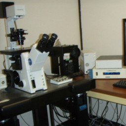
The AS MDW was developed for live cell fluorescence imaging. The system uses the Leica
DM IRE2 inverted stand with motorized objective turret, z positioning, and fluorescence
filter changing. In addition to a mercury lamp it has a Xenon lamp in a monochromator.
This latter source is more stable over time and causes less phototoxicity in comparison
to the mercury lamp.
The AS MDW also has a motorized stage so that multiple fields can be imaged, over
time, in the same experiment, increasing the number of cells analyzed. There is a
humidified chamber which fits on the stage and can be perfused with custom mixed gases,
such as 5% CO2 in air, to maintain the proper pH of bicarbonate-buffered media.
The system is equipped with N PLAN L 40x/0.55 CORR, HCX PL APO 63x/1.30 GLYCEROL and
HCX PL APO 100x/1.4 OIL objectives. Differential interference contrast optics provide
contrast in bright field images. The 63x objective is optimized for 37°C and is on
a Piezo mount for 3ms focus changes during z stack acquisition. On the bottom port
of the stand is a Photometrics Coolsnap HQ camera. Multiple image planes can be acquired
at each time point and the resulting data can be processed by 3D image deconvolution.
2 Leica DM IRE2 Microsystems
We have two Leica DM IRE2 systems, one within an environmental chamber for live cell
imaging. Features include:
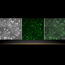
There are two of these systems, one within an environmental chamber for live cell
imaging. Each uses the Leica DM IRE2 inverted stand with motorized objective turret,
z positioning, and fluorescence filter changing. The objectives on each include 10x/0.25n.a.
Ph1, 40x/0.6n.a. Ph2 HCX PL FLUOTAR, 63x/1.4 HCX PL APO and 100x/1.4 HCX PL APO. The
microscopes use phase contrast with the 10x and 40x objectives and differential interference
contrast with the 63x objective.
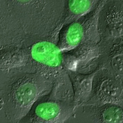
100W mercury arc lamps provide excitatory light for fluorescence imaging and there
are filter cubes for DAPI, GFP (FITC & Cy2), TRITC (Cy3 and Texas Red) and Cy5 (Alexafluor633).
Both DM IRE2 microscopes have CCD cameras for fluorescence imaging, one a Leica DC350
FX and the other a Photometrics Coolsnap HQ. The microscopes and cameras are controlled
with personal computers running Leica FW4000 software. The software enables the user
to set up image acquisitions incorporating z-series, time series and multiple color
channels. The FW4000 software also performs basic analysis and image processing functions.
3 Carl Zeiss Axiovert 200 Microscopes
We have three identically configured Zeiss Axiovert microscopes on air tables for
routine phase contrast and color imaging of live and stained specimens.
There are three identically configured Zeiss Axiovert microscopes on air tables. Each
is equipped with 10x, 20x and 32x objectives, phase contrast optics, a Zeiss AxioCam
MRc color camera and a PC interface running Zeiss Axiovision. These are used for routine
phase contrast and color imaging of live and stained specimens.
Olympus IX81 Fluorescence Microscope
The Olympus IX81 is an inverted system microscope built for live cell imaging. Features
include:
— Heated stage insert — Filter wheel for short-term live cell imaging of rapid processes
The Olympus IX81 has an inverted motorized stand controlled by the IX2-UCB control
box. Fluorescence illumination is from a Sutter Instruments Lambda LS Xenon lamp with
liquid light guide. Fluorescence filter changing is done by the Sutter Instruments
Lambda 10-3 filter changer. There are filter cubes for DAPI, FITC, and TRITC. For
more rapid filter changing for live cell imaging there is a filter wheel containing
emission filters for DAPI, FITC, TRITC and Cy5. Also for live cell imaging there is
a Bioptechs Delta T5 culture dish temperature controller. The objectives include a
UPlanFL N 10x/0.30 Ph1, a LUCPlan FL N 40x/0.60 Ph2 with correction collar for long
working distance, a UApo/340 40x/1.35 oil iris which transmits UV light for 340/380
ratio imaging, a PlanApo N 60x/1.42 oil and a UPlanSApo 100x/1.40.
Carl Zeiss LSM700 Confocal Microscope
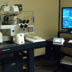
Our LSM700 laser scanning microscope is a member of the seventh generation of confocal
instruments from Carl Zeiss. Features include:
— Confocal point scanning — 3D reconstruction — Multi-field time lapse — Ratiometric concentration determination — Tile and stitch — FRET (Förster resonance energy transfer) — FRAP (fluorescence recovery after photobleaching)
The LSM700 has a scan head with two photomultiplier tube (PMT) detectors on an
inverted Axio Observer Z1 stand. There are four solid state lasers producing lines
at 405nm, 635nm, 555nm and 488nm. A transmitted light PMT collects transmitted light
images using differential. A secondary dichroic beamsplitter facilitates acquiring
lambda scans to be used for linear unmixing.
The entire microscope stage and objective turret are enclosed in an environmental
chamber to have stable temperature and CO2 control for live cell imaging. There is
a motorized stage for acquiring multiple fields in separate wells of a multiwell plate
at each time point in time lapse data acquisition.
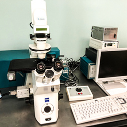
ZEISS PALM System for Laser Microdissection
ZEISS PALM system for laser microdissection combines powerful imaging capabilities
with robust and operationally simple microdissection technology.
— Applicable to cryosections, FFPE (formalin-fixed and paraffin embedded) tissue, native
tissue like fresh plants, live cells and chromosomes - without contamination — Suitable for laser microdissection and analysis for DNA, RNA and protein isolation,
whether from archive material or live cells — Works with standard glass slides
ZEISS PALM system for laser microdissection combines powerful imaging capabilities
(bright field, phase contrast, multi-channel fluorescence) with robust and operationally
simple microdissection technology.
PALM MicroBeam uses a focused laser beam to cut out and isolate the selected specimen
without contact. The patented laser catapult isolates the target area fast and uncontaminated.
It allows to obtain the homogenous analysis material necessary for meaningful scientific
results. The short laser pulse minimizes time heat transfer to adjacent areas, allowing
isolation of vital life cells.
Because analyses of gene expression patterns rely on exactly-separated analytical
material, unwanted cells may alter your results and conceal the signals of the relevant
cells. PALM MicroBeam prevents this by allowing you to define cells and tissue regions
precisely, ensuring your results are exact and reproducible.
Challenge the conventional. Create the exceptional. No Limits.